Schistocephalus solidus
LIFE CYCLE IN THE WILD
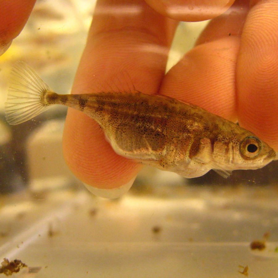
The tapeworm Schistocephalus solidus (Müller, 1776) is a trophically transmitted pseudophyllidea cestode with a three-host complex life cycle. Schistocephalus solidus can infect different species of cyclopoid copepod (as first intermediate host) and fish-eating bird (as final host), but it is highly specific to its second intermediate host, the three-spined stickleback (Gasterosteus aculeatus). It is typically found in populations of lake sticklebacks.
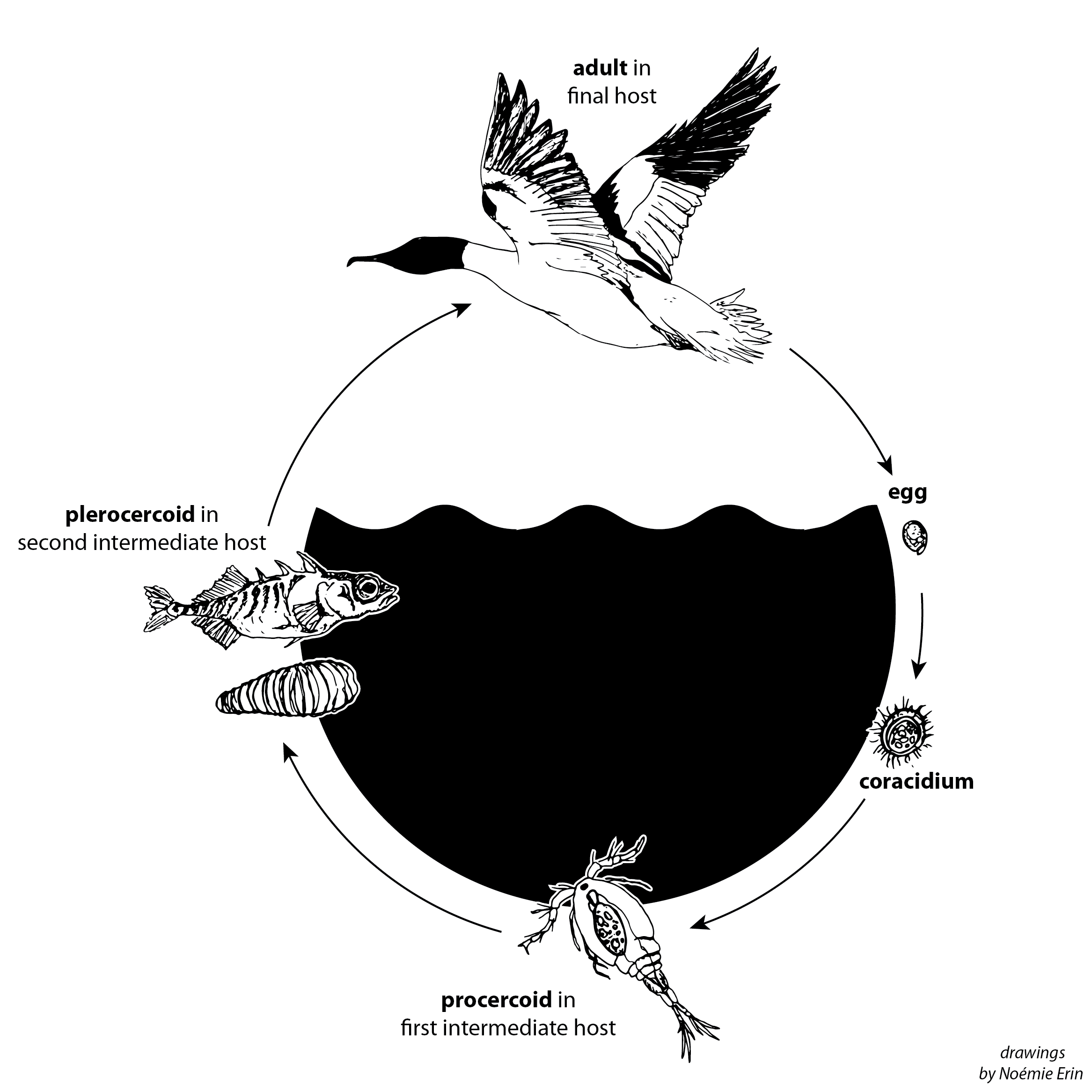
The life cycle starts when a fish-eating bird (host 3: final host) infected with mature tapeworms disperse S. solidus eggs with their faeces. After two to three weeks in freshwater, the eggs hatch and release the free-swimming larval stage, coracidium, which has to be ingested by a cyclopoid copepods (host 1: first intermediate host) within a few hours to continue the cycle. Once established in the copepod’s haemocoel, S. solidus will develop into the procercoid larval stage and become infective to the next host within a few days. When a three-spined stickleback (Gasterosteus aculeatus; host 2: second intermediate host) feeds on an infected copepod, the procercoid penetrates the gut wall to establish in the fish’s body cavity. Through this process, the procercoid shades its outer tegument and develops into a plerocercoid stage, which will grow for several weeks in its intermediate host before becoming infective to its final host. Fish-eating birds preying on infected sticklebacks will complete the cycle, allowing the simultaneous hermaphrodite tapeworms to reproduce sexually (by self- or cross-fertilization) in the bird digestive tract.
LIFE CYCLE IN THE LAB
The entire life cycle of Schistocephalus solidus can be reproduced in the laboratory§.
1) Collection
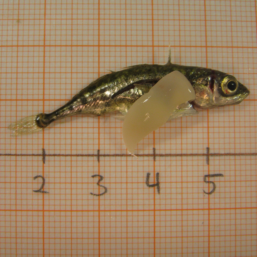
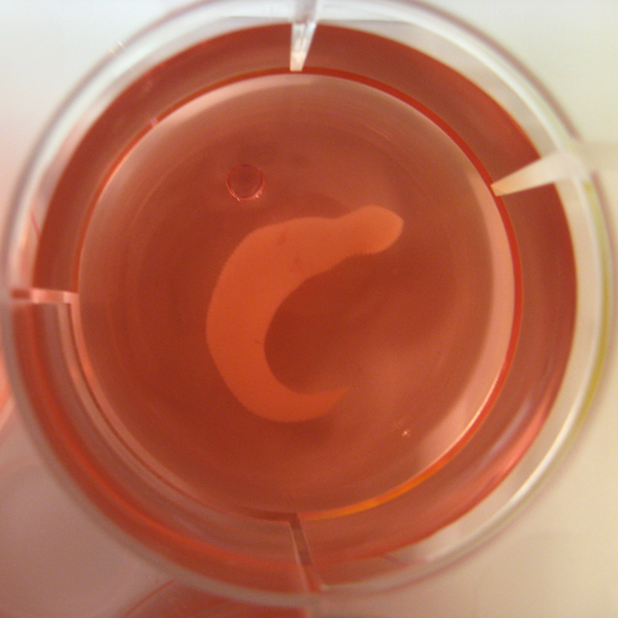
Infected three-spined sticklebacks are sampled from the wild and brought to the laboratory. Fish are killed, with an overdose of anaesthetic or by sectioning the spine, to dissect out S. solidus plerocercoids from the body cavity. The plerocercoids are then placed in medium.
2) In vitro breeding
Plerocercoids are paired and enclosed into a mesh bag. The mesh bag is secured to the cap of a medium bottle, which is placed into a dark shaking water bath at +40°C. This reproduces the conditions of a bird digestive tract and the plerocercoids start to mature, mate and produce eggs that fall at the bottom of the medium bottle. Every day for a week, the eggs are collected, washed from the medium and stored in tap water at +4°C.
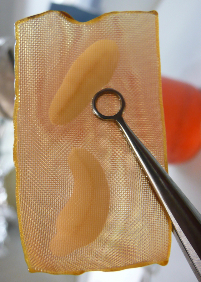
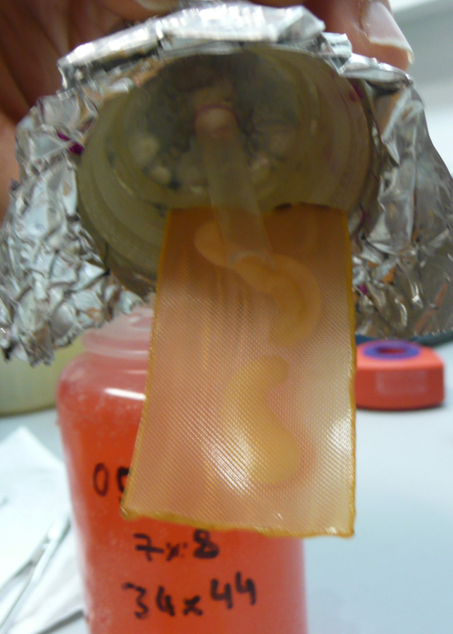
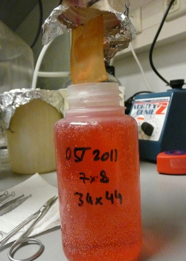
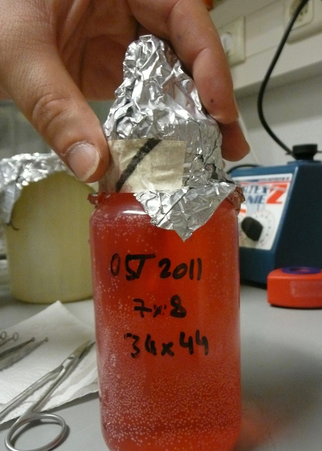
3) Copepod infection
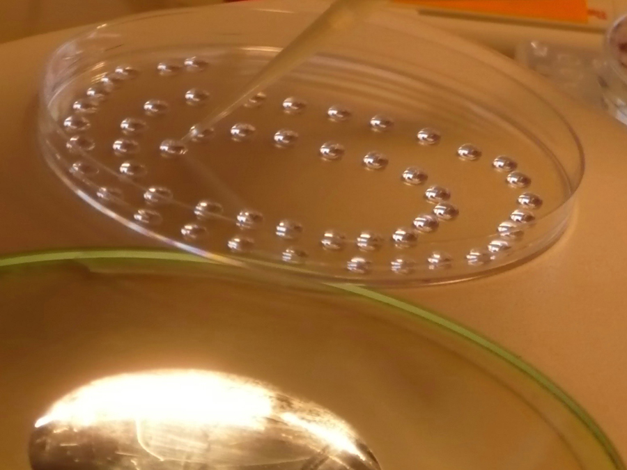
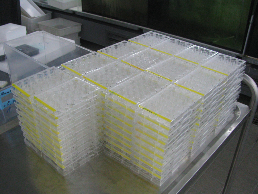
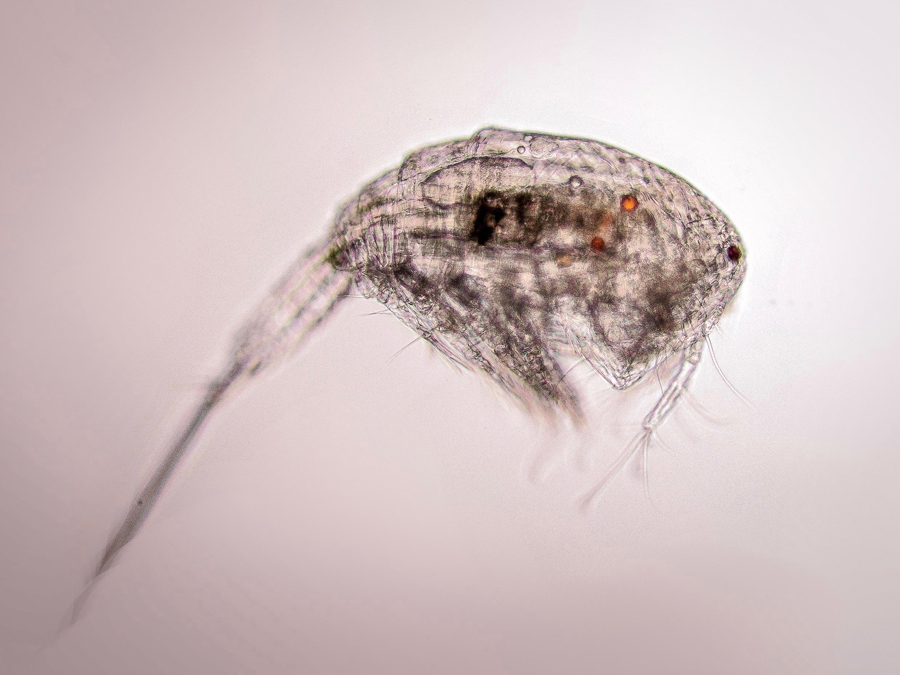
Eggs are incubated for three weeks in the dark at +20°C. Coracidia are then triggered to hatch by a change in light regime. Coracidia can then be fed to lab-bred cyclopoid copepods. Starting from four days after parasite exposure, copepods can be check under a microscope for the presence of a developing procercoid to confirm infection.
4) Fish infection
Within two weeks in the copepod, procercoids are infective to three-spined sticklebacks. Infected copepods can be fed to lab-bred fish, completing the cycle!